The time while a patient is wearing braces, rather than retainers.
Bonding paste, which is used to attach the braces to the teeth.
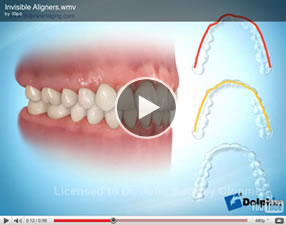
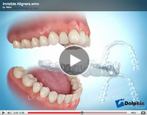
These systems all consist of a series of clear plastic aligners worn over the teeth. They are almost invisible. Each aligner is worn for 1-2 weeks before moving onto the next aligner. These systems are not suitable for every orthodontic problem. Please see invisalign aligners in appliances section.
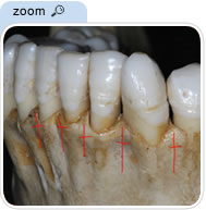
The bone surrounding each tooth.
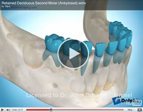
An abnormal union between two bones or between a root surface and surrounding bone resulting in tooth immobility.
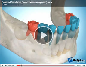
This clip shows a retained deciduous tooth , which interfered with the eruption of the permanent tooth.
Front
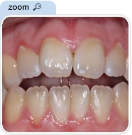
The lower incisors are not overlapped in the vertical plane by the upper incisors and do not occlude with them.
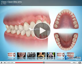
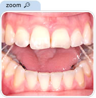
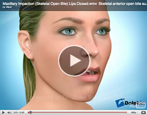
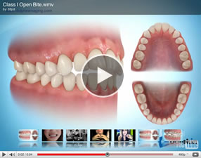
This clip shows an anterior open bite.
A wire engaged into orthodontic brackets which delivers a force to produce tooth movement.
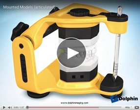
An instrument that mimics the movements of the lower jaw to which individual patients upper and lower dental study models can be attached.
To facilitate the movement of teeth with invisalign/ aligners, it is commonly necessary to place attachments on the teeth, so the aligner can grip the tooth. Please see our blog.
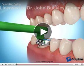
The metal ring that is cemented to a tooth for strength and anchorage.
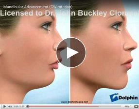
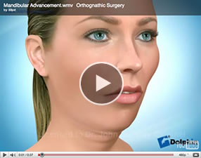
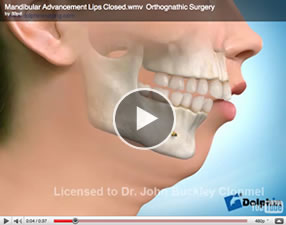
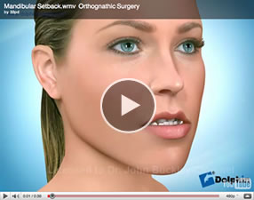
A surgical procedure to reposition the lower jaw forwards, backwards or by rotation.
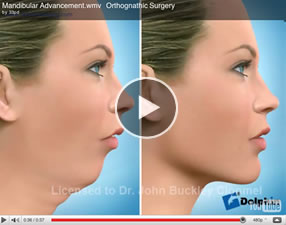
These clips show a mandibular ( lower jaw ) advancement.
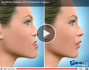
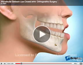
These clips show a mandibular ( lower jaw ) setback
Relating to the upper and lower dental arches or jaws.
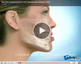
Surgery to reposition both upper and lower jaws.
This clip shows a procedure to move the maxilla ( upper jaw ) forwards, and the mandible ( lower jaw) backwards.
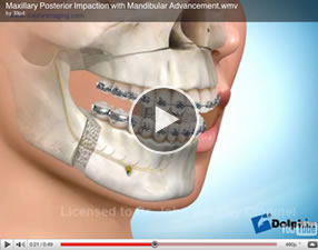
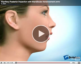
This clip shows a procedure to move the maxilla (upper jaw) upwards more so at the back, and the mandible (lower jaw) forwards.
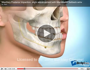
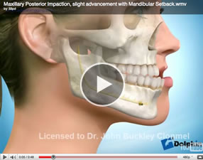
This clip shows a procedure to move the upper jaw upwards and slightly forwards and the lower jaw backwards.
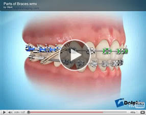
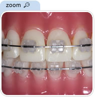
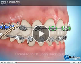
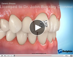
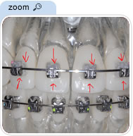
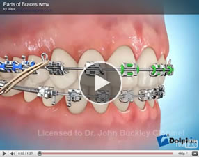
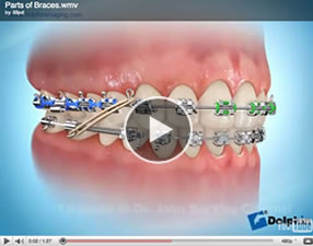
A word commonly used to describe a fixed orthodontic appliance, usually comprised of brackets, bands and wires.
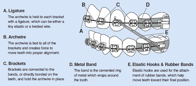
Picture Courtesy OF A.A.O.
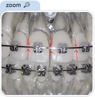
A precisely fabricated fixed orthodontic attachment made from metal, ceramic or plastic that is bonded to the teeth. The bracket has a slot that the archwire fits into.
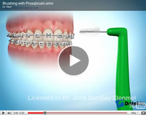
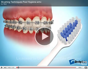
Brushing the teeth is part of an individual’s daily home dental care. Patients with braces should brush the teeth and braces for 2-3 minutes after every meal and last thing at night.
For school children they should be brushed after breakfast , upon returning from school, after the evening meal , and last thing at night.
After brushing before you spit out it is worthwhile to swish the froth of toothpaste around your teeth and mouth for about a minute, this will act somewhat like a fluoride mouth rinse . It is best to then spit out the toothpaste but not to rinse with water after brushing.
Grinding the teeth, usually during sleeping. Bruxism can cause abnormal tooth wear and may lead to pain in the jaw joints.
The cheek side of the teeth.
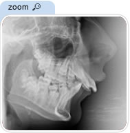
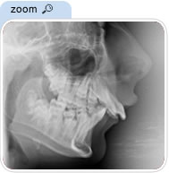
A lateral (side view) x-ray of the head.
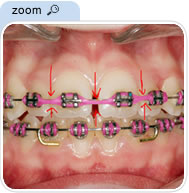
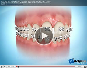
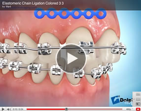
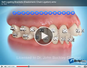
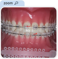
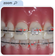
A stretchable series of elastic o-rings connected together and placed around each bracket to hold the archwire in place and move the teeth.
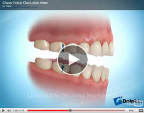
The lower incisors bite 2-4 mm behind the upper incisors. This is the ideal relationship of the teeth.
This clip shows the ideal relationship of the teeth.
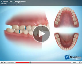
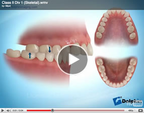
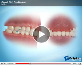
The lower incisors bite further behind the upper incisors then normal and the upper incisors are retroclined. These clips show teeth with a class 2 division 1 incisor relationship.
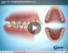
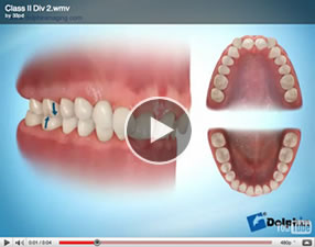
The lower incisors bite further behind the upper incisors then normal and the upper incisors are retroclined. These clips show teeth with a class 11 division 11 incisor relationship.
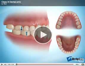
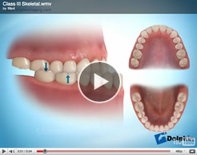
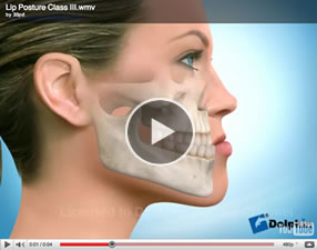
The lower incisors bite further forwards of the upper incisors than normal. These clips show teeth with a class 111 incisor relationship.
An overbite in which the lower incisors make contact with either the upper incisors or the gum tissue in the roof of the mouth.
Complete orthodontic treatment performed to correct a malocclusion.
A genetic occurrence in which the expected number of permanent teeth do not develop.
A birth defect that may occur as an isolated phenomenon or as part of a syndrome, consisting of premature fusion of one or more skull sutures.
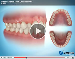
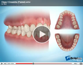
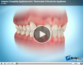
The upper incisor teeth or upper molar teeth bite on the inside on the lower teeth.
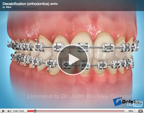
Often used to describe the loss of mineral from the tooth surface immediately surrounding an orthodontic appliance. It is caused by excessive intake of sugar and accumulation of bacteria (plaque) on the tooth surface and insufficient tooth brushing. It results in permanent discolouration of the tooth surface and cavitation if extreme.
The process of removing dental compensations, using fixed appliances, where the teeth have attempted to naturally compensate for a mismatch in the jaw relationship. Decompensation is often carried out prior to surgical correction of jaw relationships. This may involve wearing upper and lower braces for 6-12 months prior to surgery.
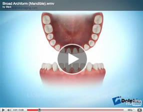
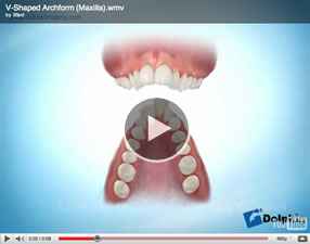
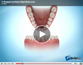
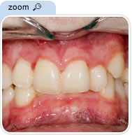
The arch formed by the upper and lower teeth when viewed from below or above respectively.
The statutory body which regulates the practice of dentistry in the Republic of Ireland. The members are appointed from the dental profession, the universities, the medical council, the government and the public.
The inclination of the teeth is naturally altered to help compensate for a mismatch in the jaw relationship.
Also known as deep overbite, this occurs when the upper front teeth overlap the bottom front teeth in a vertical direction to an excessive amount.
The material and information that the orthodontist needs to properly diagnose and plan a patient’s treatment. Diagnostic records may include a thorough patient health history, a visual examination of the teeth and supporting structures, models of the teeth, a wax bite registration, extraoral and intraoral photographs, a panoramic and a cephalometric radiograph.
A space between teeth (most commonly the upper central incisors).
Plaque disclosing tablets are a useful aid to help patients maintain their oral hygiene.

Away from the midline.
A surgical technique for lengthening bones.
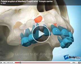
The eruption of a tooth in an abnormal position.
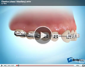
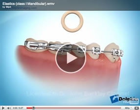
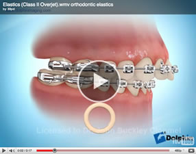
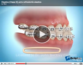
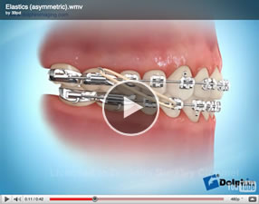
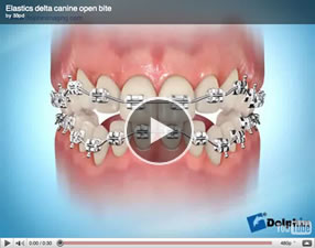
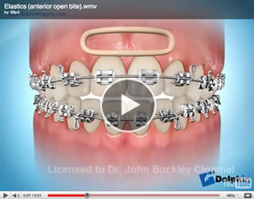
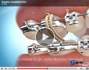
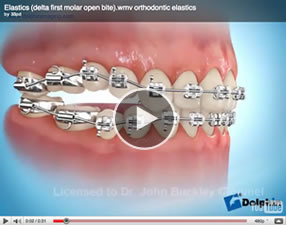
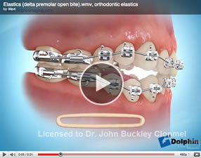
Elastics or rubber bands which maybe worn during certain stages of treatment to achieve orthodontic tooth movement.
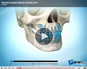
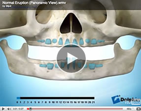
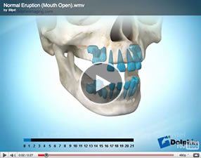
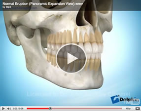
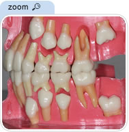
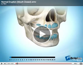
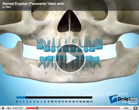
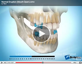
The process by which teeth enter into the mouth. The following clips show the normal eruption process. The line at the bottom of the clips indicates the patients age in years.
A clear appliance worn after braces to keep the teeth straight. You may wish to see our entry for Retainers in our Treatments section of this website.
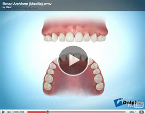
An orthodontic appliance to increase the width of the upper dental arch or jaw.
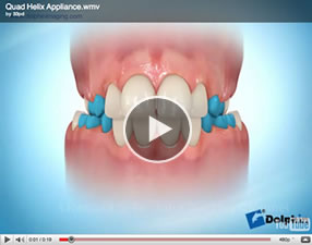
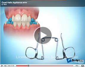
This clip shows a quadhelix which is a fixed expansion appliance. It is not removable by the patient.
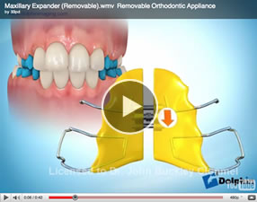
This clip shows a removable expansion appliance for the upper teeth . It is removable by the patient . The midline expansion screw is turned once a week or more by the patient or their parent.
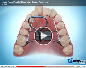
This clip shows a hyrax expansion appliance . It is not removable by the patient. The midline expansion screw is turned once or twice a day.
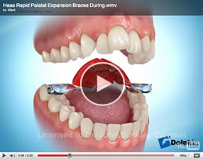
This clip shows a Haas expansion appliance . It is not removable by the patient. The midline expansion screw is turned once or twice a day.
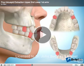
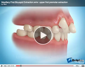
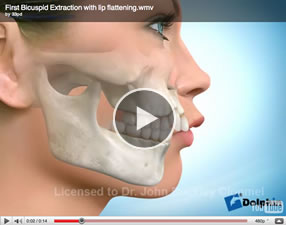
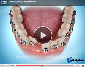
The removal of a tooth . Extraction of teeth for orthodontic reasons is something which is only done after it has been considered carefully. If an equally good result or a better orthodontic result can be achieved without extracting teeth then the decision is a “no brainer”. However in some orthodontic cases the best result can only be achieved by extracting teeth. It is important to appreciate that when teeth are extracted for orthodontic reasons that the spaces created by the orthodontic extractions will be closed at the completion of orthodontic treatment. Ultimately the patient or parent can make a decision in an informed way once their options are explained to them. These clips show various cases where extractions were used to achieve specific orthodontic objectives.
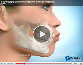
This clip shows a patient with protrusive upper and lower lips , upper teeth which are sticking out too far, and crowding in the upper and lower jaws. The orthodontic objectives are to retract the protrusive upper and lower lips, to retract the upper and lower front teeth and to correct the crowding problem. To achieve these treatment objectives four first premolars were extracted.
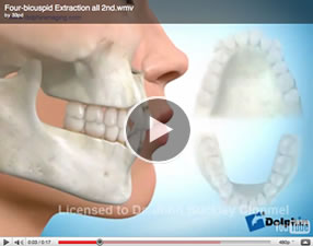
This clip shows a patient whose lips are in a good position there is crowding of the upper and lower teeth. The treatment objective is to correct the crowding.It was felt that sufficient space could not be created by other appropriate orthodontic means than extraction. To achieve this objective four second premolars were extracted.
This patient had a protrusive lower lip, lower front teeth biting in front of the upper front teeth , and crowding of the upper and lower teeth . The treatment objectives were to retract the lower lip, to retract the lower front teeth so that they would bite behind the upper front teeth as is the normal relationship and to correct the crowding problem. It was felt that these treatment objectives could not be achieved without extractions. Upper second and lower first premolars were extracted.
The following two clips describe the same case where the lower teeth are straight , but the upper lip is protrusive , the upper teeth stick out and are crowded. The treatment objectives are to create enough space to retract the upper teeth and consequently the upper lip ,and to correct the crowding of the upper teeth. In many cases which are similar to this but possibly a little less severe it is possible to create enough space in the upper jaw to achieve the treatment objectives outlined above without extractions , by using for instance headgear or a forsus type appliance amongst many other equally good appliances. It was decided here to extract both upper first premolars to achieve these objectives . Often the treatment plan reflects the patients preferences regarding which appliances they are prepared to wear.
Occasionally under specific circumstances one lower front tooth maybe extracted
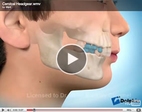
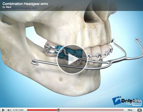
A wire appliance used with headgear. Primarily used to move the upper first molars back, creating room for crowded or protrusive front teeth. The facebow has an internal wire bow and an external wire bow. The internal bow attaches to the buccal tube on the upper molar bands inside the mouth and the outer bow attaches to the breakaway safety strap of the headgear. These three clips illustrate a facebow being worn with three different types of headgear, cervical , combination, and high pull .
Cervical headgear.
Combination headgear.
High Pull Headgear
An appliance that is fixed to the tooth surfaces in order to produce tooth movement.
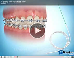
An important part of daily home dental care. Flossing removes plaque and food debris from between the teeth, brackets and wires. Flossing keeps teeth and gums clean and healthy during orthodontic treatment.
This provides a constant light force to correct a situation where the upper teeth are too far in front of the lower teeth. On certain occasions it maybe an alternative to headgear.
A fold of mucous membrane
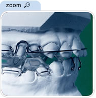
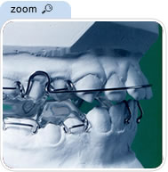
Appliances that utilize the muscle action produced when speaking, eating and swallowing to produce force to move the teeth and align the jaws. They are also known as orthopaedic appliances. The “Twin Block” and “Herbst appliance” are two well-known examples of functional appliances. You may wish to see the Functional appliances section in Treatments on this website
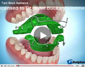
Twin block appliance.
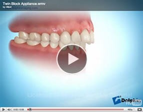
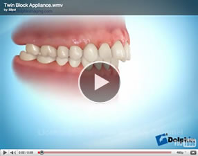
Clip of twin block appliance.
Please see video clips
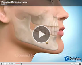
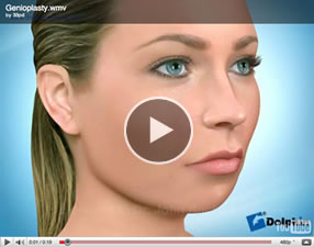
A surgical procedure to reposition the chin.
Reduction genioplasty surgery. ( chin surgery to reduce prominence of chin )
Advancement genioplasty surgery. ( chin surgery to increase prominence of a small chin)
The gum tissue surrounding teeth.
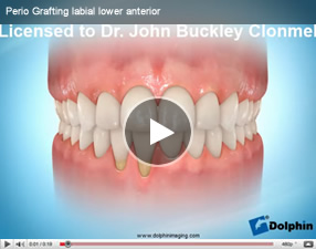
A shift of the gum margin exposing the root surface.
Any material or tissue that is not normally part of an organ or tissue, implanted or transplanted for the purpose of reconstruction or repair.
Showing an excessive amount of gingival (gum) tissue above the front teeth when smiling.
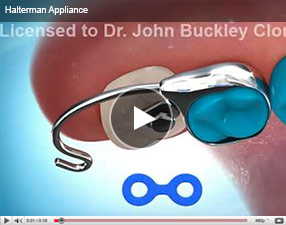
The Halterman appliance is a fixed ( not removable by the patient ) appliance, which can be used to disimpact (free up) , impacted(stuck) first permanent molars. First permanent molars normally erupt at the age of 6 years , so it is at this time that the problem may arise. If it is possible to correct the problem in a timely fashion, it may prevent more problems arising as the rest of the adult teeth erupt.
Halterman appliance
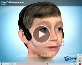
An appliance worn outside of the mouth to provide traction for growth modification and tooth movement .It is normally only worn when you are at home and in bed at night. ( please see face bow above )
These three clips illustrate a facebow being worn with three different types of headgear, cervical , combination, and high pull .
Cervical headgear.
Combination headgear.
High Pull
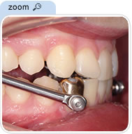
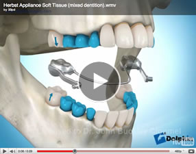
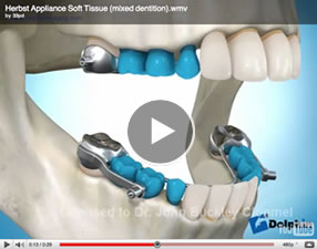
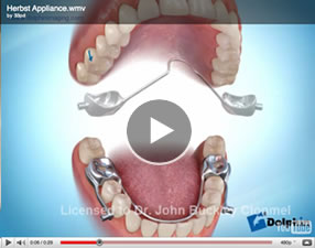
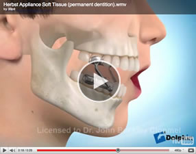
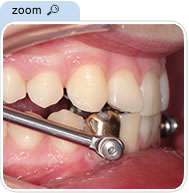
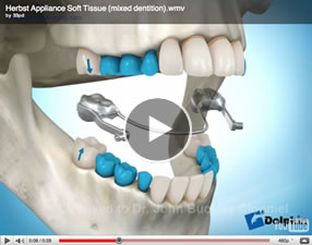
A type of functional appliance. This appliance is used to move the lower jaw forward.
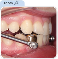
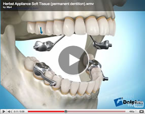
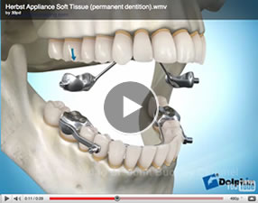
This clip shows the Herbst appliance being used in a patient who has both deciduous ( baby ) and permanent teeth.
These two clips show the Herbst appliance being used in an older patient when all the deciduous ( babyteeth) are gone and have been replaced by permanent teeth.
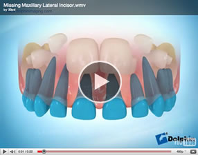
The developmental absence of teeth .
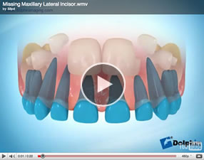
This clip shows a situation where a patient’s upper right lateral incisor fails to develop.
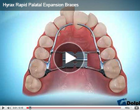
The Hyrax appliance is a fixed (not removable by the patient) expansion appliance. It is normally turned once or twice a day. It is used to achieve transverse (sideways) skeletal expansion of the upper jaw.
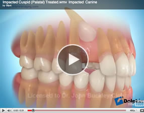
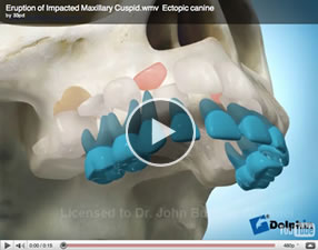
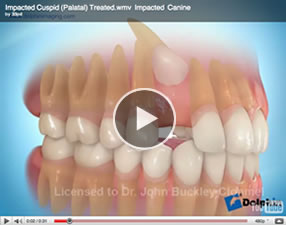
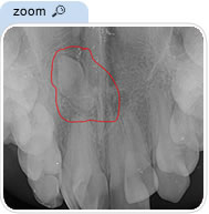
A tooth that does not erupt into the mouth or only erupts partially is considered impacted.
This clip shows an impacted canine.
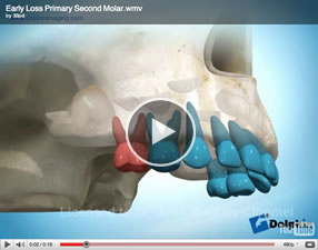
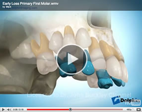
These clips show impacted upper second premolars due to early loss of the baby teeth.
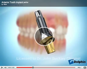
An artificial material that is placed into the body. Often used to refer to dental implants.
Please also see TAD(S) in this section
An imprint of the upper and/or lower teeth used to make study models of the teeth
With the lower jaw in the rest position the lips are apart and muscular effort is required to obtain a lip seal.
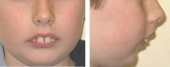
A nerve which travels in the inferior dental canal (see below) and supplies sensation to the lower teeth, the lower gum tissue and the skin covering the lower lip and chin.
A canal that runs in the lower jaw that contains the inferior alveolar (dental) nerve (see inferior alveolar nerve).
A situation in which a tooth or group of teeth are positioned below the level of adjacent teeth commonly due to ankylosis (see ‘ankylosis’).
Orthodontic treatment performed to intercept a developing problem. Usually performed on younger patients who have a mixture of primary (baby) teeth and permanent teeth. You may wish to see our blog on interceptive treatment.
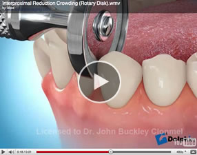
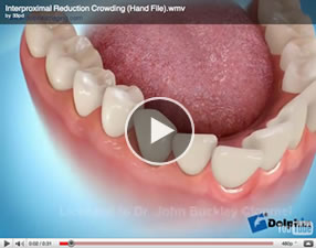
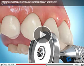
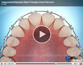
Removal of a small amount of enamel from between the teeth either to provide space for relief of crowding or to eliminate triangular gaps ( black triangles) between the teeth. Also known as reproximation, slenderizing, stripping, enamel reduction or selective reduction.
These two video clips show interproximal reduction for relief of crowding.
These two video clips show interproximal reduction to eliminate “black triangles” i.e. gaps between the teeth near the gums.
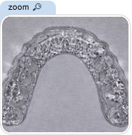
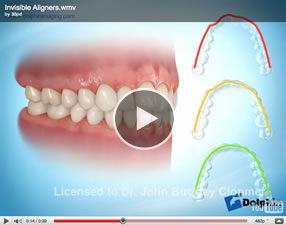
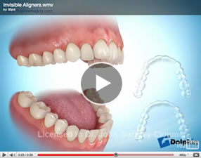
These systems all consist of a series of clear plastic aligners worn over the teeth. They are almost invisible . Each aligner is worn for 1-2 weeks before moving onto the next aligner. These systems are not suitable for every orthodontic problem. Please see Invisalign and aligners in the Treatment section of this website.
The published scientific work on invisalign however suggests that it is not a suitable appliance for treating every case if the best result is to be achieved.
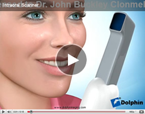
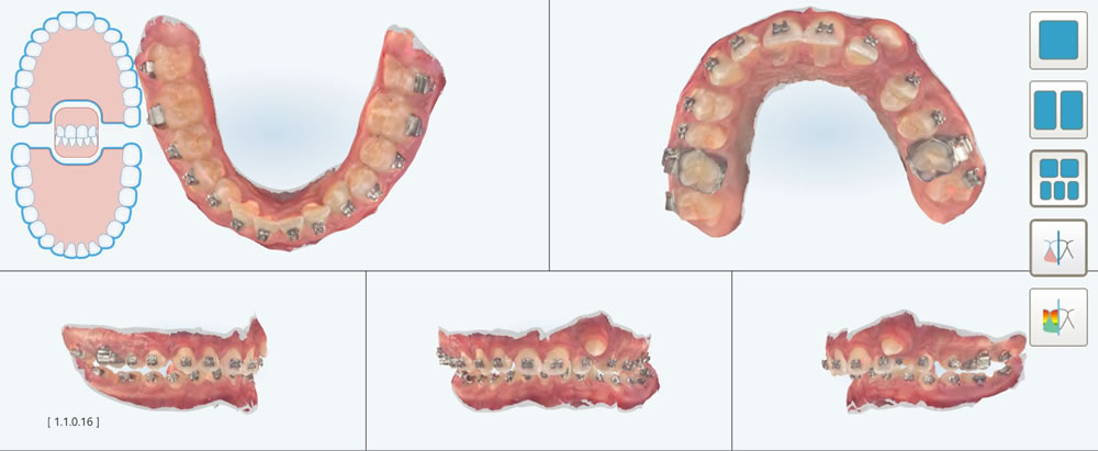
An intraoral scanner is a device for capturing direct optical impressions in dentistry. An example of one type is the iTero scanner. The iTero scanner is the one that we use in Clonmel Orthodontics.
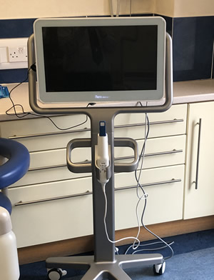
It produces a vitual image or scan of the teeth.
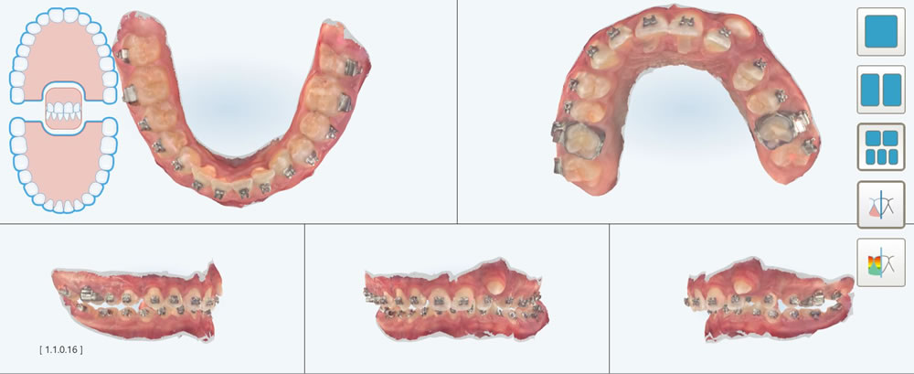
This virtual model will replace gypsum( plaster of Paris) models in certain circumstances. Also the virtual model can be manipulated to simulate orthodontic treatment.
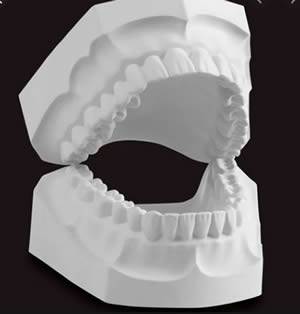
The surface of the teeth in both arches that faces the lips.
The segment of the jaw containing the incisor teeth.
The groove created where the curvature of the lower lip merges with the skin overlying the lower lip.
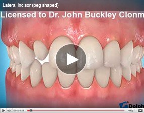
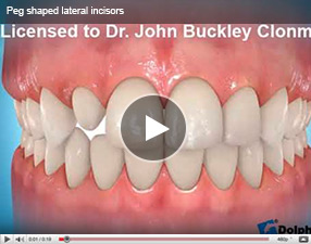
Peg shaped lateral incisors are normally smaller both in their width and height. This video shows a peg shaped lateral incisor being built up to a more normal size by the addition (bonding) of composite to the tooth.
A radiograph of the side of the face and skull taken in a standardised reproducible position.
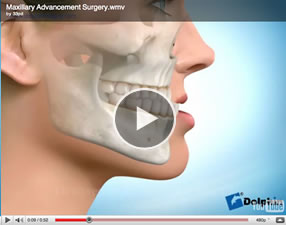
A surgical procedure of the tooth bearing segment of the upper jaw used to reposition the jaw most commonly in an anterior, upward (impaction) or downward direction.
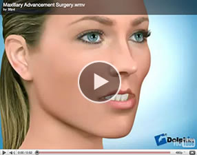
Upper jaw advancement surgery.
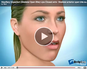
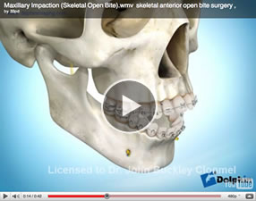
Upper jaw moved vertically
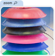
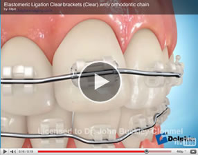
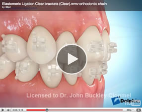
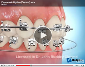
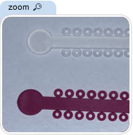
A small elastic o-ring, shaped like a doughnut, used to hold the archwire in the bracket.
Please see these video clips
Coloured and clear Elastic o- rings
The tongue side of the teeth in both arches.
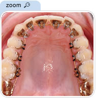
Orthodontic braces worn on the inside of the teeth.
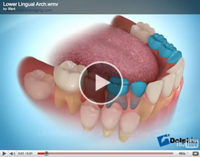
A form of fixed orthodontic appliance that runs between the inner surfaces of the lower molar teeth designed to help prevent forward movement of the molars.
Please see video clip.
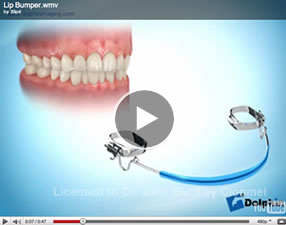
A somewhat controversial appliance it is used to attempt to move the lower molars back and possibly the lower front teeth forward, creating room for crowded front teeth. The lip bumper is an internal wire bow that attaches to the buccal tubes on the cheek side of the lower molar bands inside the mouth. The front portion of the bow has an acrylic pad or bumper that rests against the inside of the lower lip. The lower lip muscles apply pressure to the bumper creating a force that it is suggested by some orthodontists moves the molars back. It is controversial as to exactly what it achieves.
Please see this video clip
The inability to close the lips together at rest, usually due to protrusive front teeth or excessively long faces.
A poor relationship between the upper and lower dental arches or abnormal tooth positions.
The lower jaw.
The upper jaw.
The is a chamber present in the upper jaw and closely associated with the roots of the upper molar teeth.
Towards the midline.
Abnormally small jaw size.
The dental developmental stage in children (approximately ages 6-12) when they have a mix of primary (baby) and permanent teeth.
The following clips show the normal eruption process. The line at the bottom of the clips indicates the patients age in years.
A removable device used to protect the teeth and mouth from injury caused by sporting activities. The use of a mouthguard is important also for orthodontic patients.
A form of fixed orthodontic appliance that runs between the inner surfaces of the upper molar teeth designed to help prevent forward movement of the molars.
Please see video clip.
The angle formed by the junction of the nasal base and upper lip.
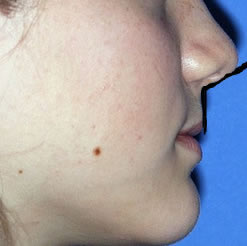
A removable appliance worn at night to help an individual minimize the damage or wear while clenching or grinding teeth during sleep
A malocclusion in which the teeth do not make contact with each other. With an anterior open bite, the front teeth do not touch when the back teeth are closed together. With a posterior open bite, the back teeth do not touch when the front teeth are closed together.
Please see video clip.
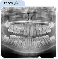
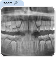
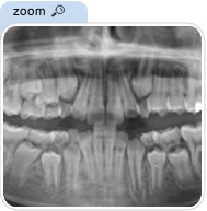
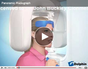
An x-ray that shows all the teeth and both jaws on one film.
A type of aligner, these systems all consist of a series of clear plastic aligners worn over the teeth. They are almost invisible . Each aligner is worn for 2-3 weeks before moving onto the next aligner. These systems are not suitable for every orthodontic problem. Please see aligners in appliances section.
Often a misleading term as the practitioner involved may not be an orthodontist . It does not mean that the practitioner has any recognised postgraduate training or qualification in orthodontics. . It merely means that the practitioner has chosen to no longer perform other general dental procedures like fillings , extractions and dentures etc.
The full list of registered orthodontists in Ireland is available from the Dental Council of Ireland (the regulatory body for dentists in Ireland) on their specialist register at http://www.dentalcouncil.ie/specialistregisters.php
Often a misleading term as the practitioner involved may not be an orthodontist . It does not mean that the practitioner has any recognised postgraduate training or qualification in orthodontics. It merely means that the practitioner has chosen to no longer perform other general dental procedures like fillings , extractions and dentures etc.
The full list of registered orthodontists in Ireland is available from the Dental Council of Ireland (the regulatory body for dentists in Ireland) on their specialist register at http://www.dentalcouncil.ie/specialistregisters.php
The specialty area of dentistry concerned with the diagnosis, supervision, guidance and correction of malocclusions. The formal name of the specialty is orthodontics and dentofacial orthopaedics.
A specialist in the diagnosis, prevention and treatment of dental and facial irregularities . Only those who have completed the requirements for specialisation of the Irish Dental Council may call themselves an “orthodontist”.
The full list of registered orthodontists in Ireland is available from the Dental Council of Ireland (the regulatory body for dentists in Ireland) on their specialist register at http://www.dentalcouncil.ie/specialistregisters.php
Also known as a functional appliance it is designed to guide the growth of the jaws and face. Please see functional appliance
Twin Block Appliance
Herbst Appliance
Surgical procedure involving cutting the bone. Which is used to facilitate moving one or both jaws surgically. You may find the following video clips to be informative.
Lower jaw advancement surgery
Lower jaw setback surgery
Upper jaw advancement surgery.
Upper jaw moved vertically
Upper jaw moved vertically and segmented
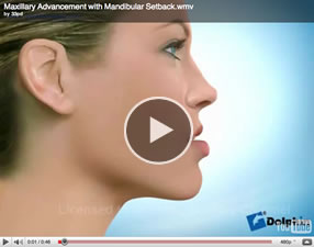
Upper and lower jaw surgery. The upper jaw is moved forwards, the lower jaw is moved back.
The upper jaw is moved upwards and the lower jaw is moved forwards.
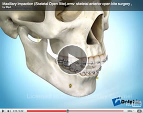
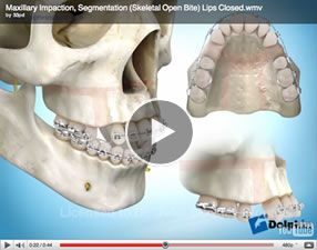
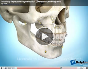
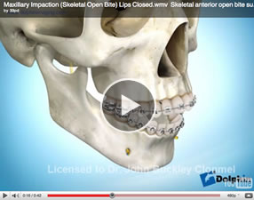
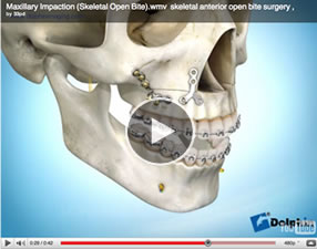
Surgical correction of an open bite.
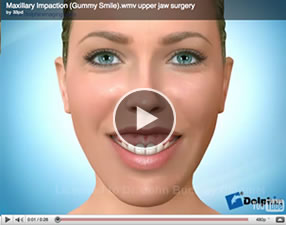
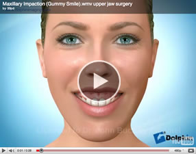
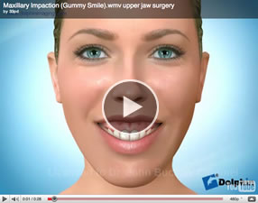
Surgical correction of a gummy smile.
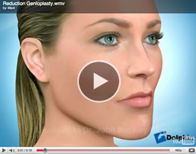
Reduction genioplasty surgery. ( chin surgery to reduce prominence of chin )
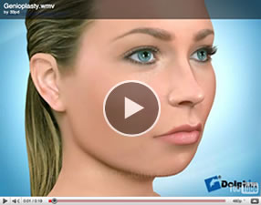
Advancement genioplasty surgery. ( chin surgery to increase prominence of a small chin)
The overlap of the lower incisors by the upper incisors in the vertical plane. Normally the upper incisor covers one third of the lower tooth .
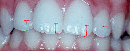
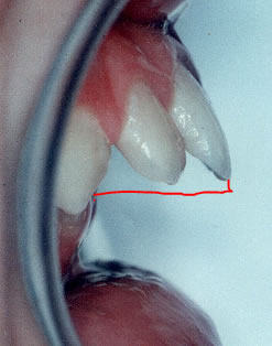
The distance between the front surface of the lower incisors and the front surface of the upper incisors. The front surface of the upper incisors is usually 2-4 mm ahead of the front surface of the lower incisors.
An x-ray that shows all the teeth and both jaws on one film.
Peg shaped lateral incisors are normally smaller both in their width and height. This video shows a peg shaped lateral incisor being built up to a more normal size by the addition (bonding) of composite to the tooth.
Diminished or abnormal sensation, such as burning, prickling, tingling or numbness.
Refers to the hard and soft tissue, or supporting structures, around the teeth.
A soft, thin film of adherent deposit on the tooth surface composed of bacteria, bacterial products and salivary constituents. Some bacteria within plaque are associated with dental decay and gum disease.
Plaque disclosing tablets are a useful aid to help patients maintain their oral hygiene.

The most anterior point on the contour of the chin.
Back.
Often a misleading term as the practitioner involved may not be an orthodontist . It does not mean that the practitioner has any recognised postgraduate training or qualification in orthodontics. It merely means that the practitioner has chosen to no longer perform general dental procedures like fillings , extractions and dentures etc.
The full list of registered orthodontists in Ireland is available from the Dental Council of Ireland (the regulatory body for dentists in Ireland) on their specialist register at http://www.dentalcouncil.ie/specialistregisters.php
Orthodontic treatment to prevent or reduce the severity of a developing malocclusion (bad bite) or crooked teeth.
The upper or lower incisors are inclined forwards to a greater degree than normal.
Protrusion of the jaw, most commonly the lower.
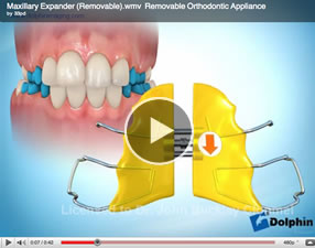
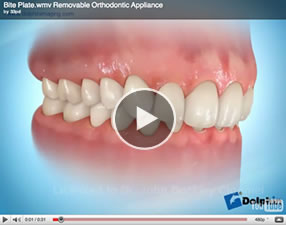
An orthodontic appliance that can be removed from the mouth by the patient. Strictly speaking of course it includes “aligners” and removable functional appliances like the “Twin block” appliance (as listed in this glossary). The term “removable appliance” also includes removable appliances which are not functional appliances (like the Twin block appliance) or aligners. Examples of these are demonstrated in these 3 video clips listed below. The appliances demonstrated below are used less commonly nowadays than they were in the past. This is because a similar treatment objective is often achieved more effectively using fixed (non-removable) appliances.
Please view these video clips
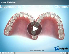
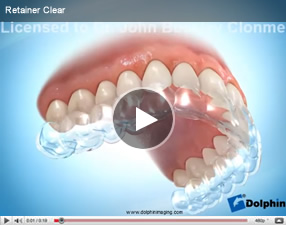
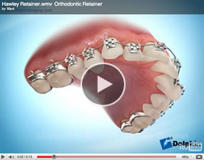
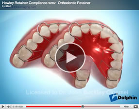
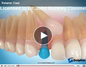
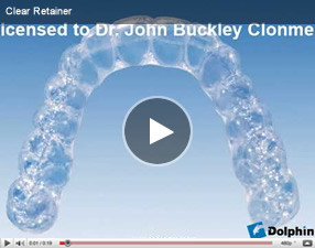
An orthodontic appliance used following orthodonic treatment in order to maintain the corrected positions of the teeth. They can be divided into bonded or fixed retainers ( which as the name suggests are bonded into place and remain in place 24 hours a day) , or removable retainers which are of the clear type or a variation of a Hawley or spring type retainer as demonstrated below
Please view these video clips
The upper or lower incisors are inclined or facing backwards to a greater degree than normal.
Retrusion of the jaw. Often used to refer to the upper and lower jaw.
Loss of root length, this often accompanies routine orthodontic treatment and is generally but not always of minor extent and of little or no clinical significance. A more severe type of resorption can occasionally be seen when an unerupted ectopic (out of normal position) tooth contacts the root of another tooth. This (the latter) type of resorption is not caused by the orthodontic movement of teeth.
Elastics or rubber bands which maybe worn during certain stages of treatment to achieve orthodontic tooth movement.
An intraoral scanner is a device for capturing direct optical impressions in dentistry. An example of one type is the iTero scanner. The iTero scanner is the one that we use in Clonmel Orthodontics.

It produces a vitual image or scan of the teeth.

This virtual model will replace gypsum( plaster of Paris) models in certain circumstances. Also the virtual model can be manipulated to simulate orthodontic treatment.

One or more of the upper molar teeth bite outside to the lower molar teeth.
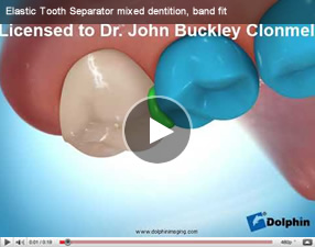
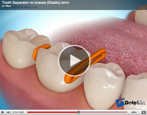
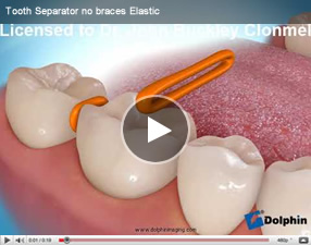
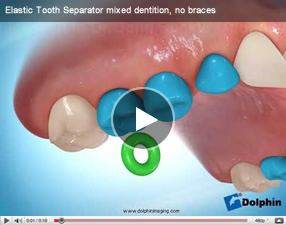
An elastic o-ring placed between the teeth to create space for placement of bands. Separators are usually placed between the teeth a week before bands are scheduled to be cemented to the teeth.
Please see these videos.
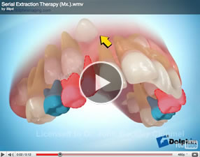
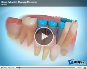
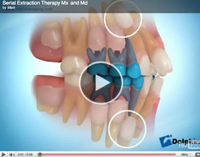
Selective or guided removal of certain primary (baby) teeth and/or permanent teeth over a period of time to create room for permanent teeth. This procedure is rarely, if ever, practised nowadays as the results achieved are often unsatisfactory, and it involves an excessive number of extractions.
Please see these videos.
These aligner systems all consist of a series of clear plastic aligners worn over the teeth. They are almost invisible . Each aligner is worn for 1-2 weeks before moving onto the next aligner . These systems are not suitable for every orthodontic problem. Please see aligners in appliances section
The relationship between the upper and lower jaws.
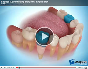
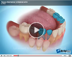
A fixed appliance used to hold space for an unerupted permanent tooth after a primary (baby) tooth has been lost prematurely, due to accident or tooth decay.
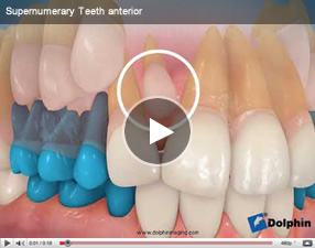
Teeth in excess of the usual number. Occasionally supernumerary teeth may interfere with the eruption of unerupted teeth , or may interfere with the orthodontic movement of erupted teeth.
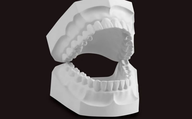
Models of the upper and lower teeth used for planning and monitoring the patients treatment.
They can be real models or virtual models as taken on our iTero scanner.
Tads or Temporary Anchorage Devices are known as mini-implants. They are used to provide anchorage to control the movement of teeth in certain situations. Please see the treatments section of this website.
An individual’s tongue pushes against the teeth when swallowing. Forces generated by the tongue can move the teeth and bone and may lead to an anterior or posterior open bite.
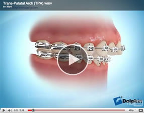
A bar which crosses the roof of the mouth and is used to stabilise or to derotate the upper back teeth.
Please see video clip.
The Twin Block is a type of functional appliance. Please see functional appliances in the Treatments section of this website section. If you wish to see details of a patient treated with a functional appliance please see.
Please see video clip.


